Fallopian tube obstruction
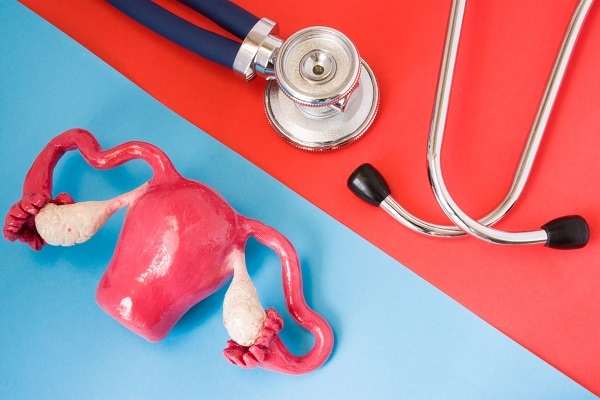
What are fallopian tubes?
The Fallopian tubes are organs specialized in the transport of gametes. They also transport and house the embryo in the first four days, providing it with nutrients, oxygen, and other conditions needed for healthy development until it reaches the interior of the uterus (endometrium), where implantation and subsequent development will take place from day five until the end of the pregnancy.
How do fallopian tubes work?
They have a sticky end (the fimbria) that actively moves to look for the egg that must leave one of the two ovaries each month. When they find it, they affix it and take it to the interior of the tube, where fertilization will take place in a region called the ampullary region. This is where the sperm must swim to meet the egg, and the Fallopian tubes serve as a highway to guide them to this meeting.
The Fallopian tubes also can move, as the intestine does, and have some cilia in their interior that are like small villi that cover it internally and have the capacity to move. By combining these two functions (peristalsis and cilia movements), we have an organ capable of moving and transporting everything inside. When fertilization occurs, the embryo is transported very effectively to the uterine cavity.
What are the most common fallopian tube diseases?
Any situation that alters the functions of fimbria, peristalsis, and ciliary motility may represent a tubal disease. This can occur because of some inflammatory disease of the pelvis or secondary to the scarring generated when a woman is recovering or fighting this type of inflammation.
Endometriosis is a typical example of pelvic inflammatory disease, as are uterine and tubal infections and surgeries such as a complicated appendectomy or surgeries for treating ovarian cysts.
In what way is the function of the fallopian tubes evaluated?
A test for Fallopian tube patency can be performed to determine the actual condition of the tubes. The most common is an imaging x-ray called hysterosalpingography, in which a catheter is passed through the cervix to administer a contrast medium that perfectly shows the uterine cavity and the path of the tubes. By taking the x-ray at different moments while administrating the contrast medium, the doctor can evaluate how the contrast medium is being transported and interpret how the tubes and the fimbria are functioning.
A similar study can also be done not with radiography but with an ultrasound. An ultrasound can evaluate the liquid content of the structures very well. Stopping liquid flow through the uterus and Fallopian tubes allows the specialist to assess how fluid flows through the uterine cavity and tubes in real time.
Another way to evaluate them is to visualize them directly through a diagnostic laparoscopy surgery. In this procedure, a contrast medium can also be administered through the cervix so that the surgeon can see exactly how the tubes behave and if they allow fluid passage without alterations.
When a woman has an obstruction in the Fallopian tubes, the problem is not necessarily accompanied by any symptoms or abnormalities that indicate that she may have issues in this part of her body. However, the doctor may suspect it based on the patient’s medical history and background and request this study to diagnose the problem and its most effective treatment.
What treatment is needed for fallopian tubes?
When the tubal obstruction is in a single location (e.g., tubal ligation) or there are external adhesions, such as appendicitis or peritonitis, it is possible to correct the problem with tubal surgery.
On the contrary, when the inflammation and damage have been to the interior of the tubes and the cilia-bearing cells have been damaged, the probability of obtaining a pregnancy is exceptionally low with corrective surgery, and the best treatment option is in-vitro fertilization.
In in-vitro fertilization, eggs are removed and fertilized with sperm in the laboratory. The fertilized eggs are then introduced into the uterus through the vagina, bypassing the Fallopian tube.